


Types of Veterinary Cystoscopy
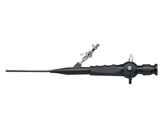
PIE-C01 Veterinary Cystoscopy
It enables minimally invasive visualization of the urinary tract, allowing precise diagnosis of bladder lesions, stone retrieval, and tumor assessment while minimizing surgical trauma and recovery time.
Veterinary canine cystoscopy Clinical Procedure Steps
Preparation:
Power on imaging system and connect cystoscopy cat to light source/video processor.
Administer anesthesia and position patient in lateral recumbency for urethral access.
Device Insertion:
Lubricate and gently advance cystoscope (≤2.7mm diameter) transurethrally.
Navigate past urethral sphincters using smooth handle articulation.
Real-time Visualization:
Monitor bladder mucosa integrity, stones, or tumors via HD display.
Capture video/images of lesions or structural abnormalities during irrigation.
Diagnosis & Documentation:
Identify pathologies (e.g., cystitis, polyps, neoplasia) on screen.
Annotate findings and generate electronic records via integrated software.
Benefits of a cystoscopy is a visual examination of the bladder
Minimally Invasive
Slim design via natural orifices.
3D Imaging
Stereoscopic bladder wall visualization.
Sterilization
Heat-resistant up to 135°C.
Precision Control
Bi-directional bending, no dead angles.
Data Management
One-touch storage of exam data.
For more information about cost of cystoscopy for dogs and buy industrial endoscope, please feel free to contact us!
If you are looking for a reliable endoscope company, please contact us.
Loading comments...